Inventions

Assays, instruments, and software.

I developed or co-developed four assays and instruments and two software tools for research questions that existing methods could not answer. A patent application has been drafted for the PLD activity assay.

Live-Cell Real-Time Phospholipase D (PLD) Activity Assay under Mechanical Force01

Assay · Patent Drafted

Live-Cell Real-Time Phospholipase D (PLD) Activity Assay under Mechanical Force

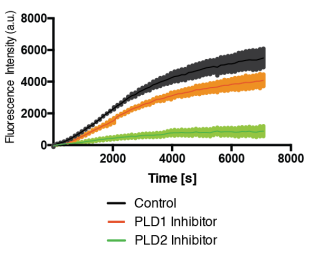
A continuous, live-cell enzyme-coupled fluorescent assay (Amplex Red / HRP / choline oxidase) that quantifies phospholipase D (PLD1/PLD2) catalytic activity in real time. The assay supports orbital shear-stress delivery in 96-well microplates, osmotic swelling, pharmacological inhibition, and volatile anesthetic exposure.

  • Real-time continuous fluorescent readout of lipid signaling kinetics
  • Adapted for in-plate mechanical stimulation (shear stress & osmotic stretch)
  • Deployed across 5 peer-reviewed publications as primary functional readout
  • High-throughput format for both enzyme readout and mechanical stimulation
View related paper
Temperature-Controlled Rapid Shear Fixation02

Instrument · Published Method

Temperature-Controlled Rapid Shear Fixation

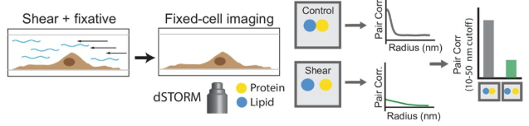
A custom parallel-plate system delivers laminar fluid shear stress while an in-line precision heater prevents thermal phase-transition artifacts. A dual PFA/glutaraldehyde injection protocol fixes mechanically stimulated nanodomain states in under 10 seconds.

  • Temperature clamping within ±0.1 °C to eliminate thermal lipid phase confounds
  • Chemically traps non-equilibrium stimulated membrane states in under 10 seconds
  • Preserves spatial organization of nanoscale lipid rafts and ion channel clusters
  • Enables post-fixation super-resolution localization of activated states
View related paper
03

Imaging · Published Method

Sub-Second Live-Cell 3D-dSTORM Super-Resolution

High-speed 3D single-molecule localization microscopy (dSTORM/SMLM) records at 200 fps (5 ms exposure) and reconstructs video at ~250 ms per frame. It captures nanodomain assembly, disassembly, and lateral translocation under shear force in live cells; traditional STORM reconstruction can require minutes per frame.

  • ~250 ms effective reconstructed video frame rate for live-cell SMLM/dSTORM
  • 200 fps acquisition camera settings on biplane 3D super-resolution optics
  • Direct visualization of lipid raft disruption and mechanosensory translocation
View related paper
VAAPR Anesthetic Sensitivity Assay Rig04

Instrument · Published Rig

VAAPR Anesthetic Sensitivity Assay Rig

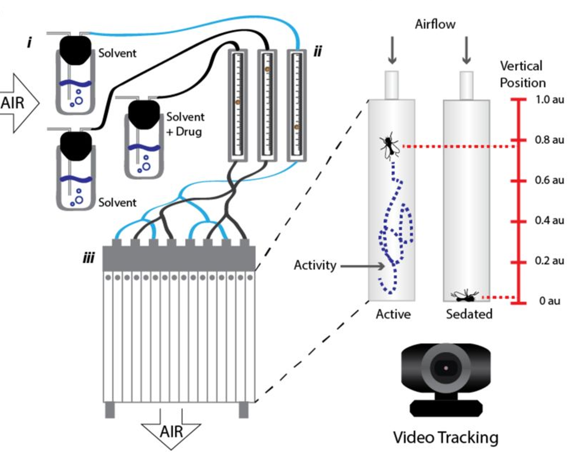
Volatile Anesthetic Administration in Parallel in Drosophila (VAAPR) combines a narrow vertical chamber array, flow-controlled vapor delivery, and automated animal tracking to calculate T50 anesthetic sensitivity across genetic variants.

  • Narrow vertical glass chamber array designed for precise gas delivery
  • Flow-controlled volatile delivery system for chloroform, isoflurane, and diethyl ether
  • Machine-vision positional recording for objective T50 determination
  • Published in bioRxiv 2019 and used in a 2020 PNAS anesthesia study
View related paper
ClockWork Circadian Behavior Analysis Suite05

Software · Private / Open Source

ClockWork Circadian Behavior Analysis Suite

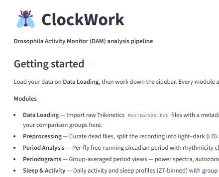
I co-developed an open-source modular Python package with an interactive Streamlit GUI for high-throughput circadian activity time-series analysis. It calculates Lomb-Scargle and chi-square periodograms, performs wavelet decomposition, and scores rhythmicity.

  • Modular Python engine for TriKinetics / DAM circadian activity data
  • Interactive Streamlit web GUI for real-time visualization and filtering
  • Automated periodogram calculation, period determination, and arrhythmicity scoring
  • NetCDF allows for simple sharing of data and analysis parameters between researchers for publication
Microscopy Training Simulator06

Software · In Development

Microscopy Training Simulator

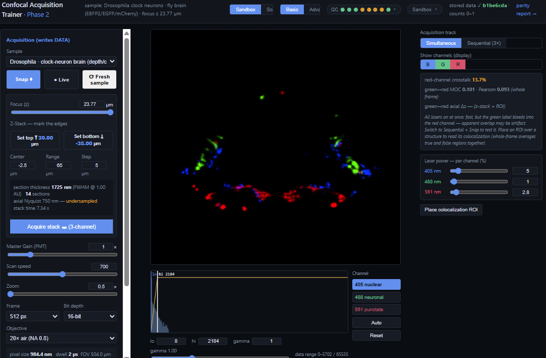
This browser-based simulator models how fluorophore brightness, photobleaching, and background interact with acquisition settings. Users can adjust pinhole diameter, channel acquisition mode, z-stack spacing, and depth across models of a cell monolayer, C. elegans, and a Drosophila brain.

  • Physics-based fluorophore model: brightness, photobleaching, background
  • Acquisition parameters change the resulting image: pinhole diameter, simultaneous vs sequential channels, z-stack spacing and depth
  • Teaches the trade-off behind each setting rather than a recipe to follow
  • Built from a decade of hands-on super-resolution and confocal experience
  • In active development